EMS12780
Bridging the gap between light and electron microscopy.
Fluorophore-Infiltrated Resin Microscopy (FIRM) is a novel staining method providing ultra-high contrast imaging of tissue subcellular structure using a standard widefield fluorescence microscope. FIRM images closely resemble those obtained by transmission electron microscopy (TEM) at low magnifications. The key concept is the infiltration of thin resin sections with a fluorophore. FIRM can be used either in epoxy or acrylate resins. When illuminated by epifluorescence, the brightly fluorescent resin reveals cell and tissue structures in negative relief, according to the light absorbing and scattering properties of such structures. The method provides much greater contrast of fine subcellular structures than obtained by typical semithin section brightfield stains, such as toluidine blue. FIRM images of multiple (>50) serial semithin sections of complex structures, such as kidney glomeruli, are easily rendered in 3-D by open source software programs.
General Methods
During the testing of a large number of fluorescent dyes as candidate counterstains for thinsection immunofluorescence in LR white sections, one with special properties was encountered. Most dyes, such as sodium fluorescein, non-selectively stained tissue and provided little contrast (not shown). However, one such dye, when applied to either LR White or Epon sections, provided very high contrast images using standard fluorescence microscopy, with the resin appearing brightly fluorescent and tissue structures appearing primarily in negative relief (Figure 1).

FIGURE 1: Human anterior pituitary, FIRM, rhodamine channel. Note that the fluorophore illuminates the LR White resin, providing an image of tissue structure primarily in negative relief. Bar, 100µm. All subsequent FIRM images are shown in monochrome.

FIGURE 2: Human anterior pituitary, formalin-fixed autopsy specimen, comparison of H&E paraffin section (left hand side) with FIRM (right hand side). 20x dry objective used for both. Scale bar, 20µm

FIGURE 3: Human anterior pituitary, FIRM image taken with 150X glycerin objective. Individual secretory granules are easily visualised, ranging from 150- 350nm in diameter. Scale bar, 5µm.
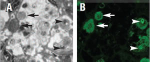
FIGURE 4: Correlative FIRM/fluorescent lectin labelling of human pituitary. A single LR White thin section was labeled first with Concanavalin AAlexa-488 (B) imaged, then stained for FIRM imaging (A). Densely granulated cells (arrows in A) are strongly labeled with Con-A (arrows in B). Dark structures resembling large lysosomes or residual bodies (arrowheads in A) are also strongly labeled by Con-A, consistent with reports in the literature.


FIGURE 5: Comparison of toluidine blue staining (top) to FIRM (left) on near adjacent sections of human kidney. Note enhanced contrast, especially of glomerular basement membranes in FIRM image. High power FIRM image (C) of glomerulus. Note resolution of podocyte foot processes (arrows). Scale bars in A, B, 20µm, in C, 5µm.
DISCLOSURE: James W. Mandell, MD, PhD, Dept. Pathology (Neuropathology), University of Virginia School of Medicine, Charlottesville, VA has filed a provisional patent relating to this invention: U.S. Provisional Patent Application Serial No. 61/298,759 Filed on January 27, 2010 Title: Compositions and Methods for Enhanced Specimen Contrast
Was this article helpful?
That’s Great!
Thank you for your feedback
Sorry! We couldn't be helpful
Thank you for your feedback
Feedback sent
We appreciate your effort and will try to fix the article